44 structure of the heart without labels
CH. 20 Assessment Flashcards | Quizlet Correctly label the following external anatomy of the anterior heart. Place the components of the electrical conducting system in order from the initiation of an action potential until the end. 1. SA node 2. Atrial myocardium 3. AV node 4. AV bundle 5. Right and left bundle branches 6. Purkinje fibers › en › healthy-livingCarbohydrates | American Heart Association Apr 16, 2018 · Carbohydrates are either called simple or complex, depending on the food’s chemical structure and how quickly the sugar is digested and absorbed. The type of carbohydrates that you eat makes a difference – Foods that contain high amounts of simple sugars, especially fructose raise triglyceride levels.
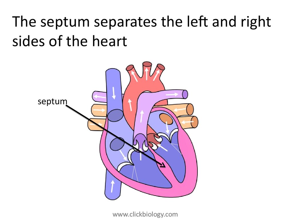
A Labeled Diagram of the Human Heart You Really Need to See The wall of the heart can be divided into three layers; outer epicardium, inner endocardium and middle myocardium. The myocardium, which consists of the cardiac muscle tissues, is responsible for the contraction of heart chambers for pumping of blood. Its contraction and relaxation leads to the heartbeat, we all are familiar with.

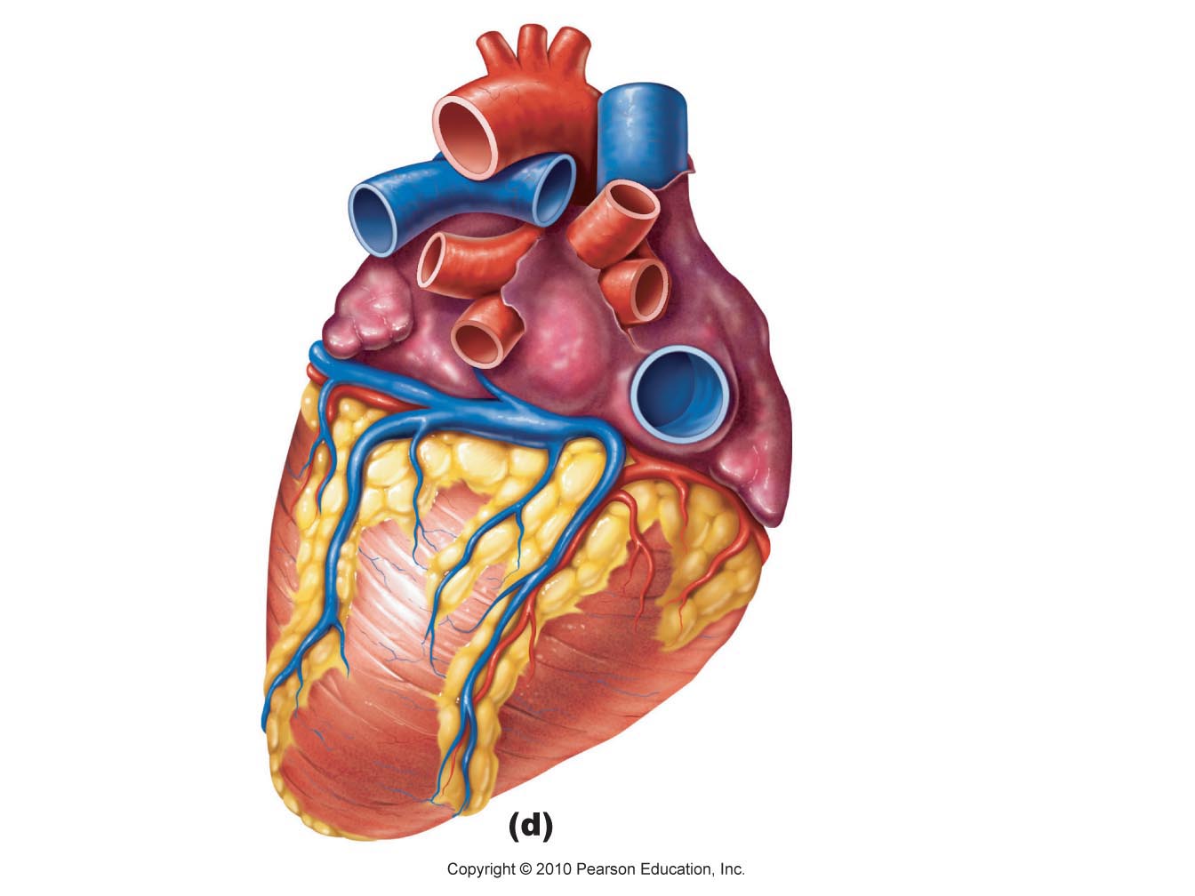
Structure of the heart without labels
Human Heart - Diagram and Anatomy of the Heart - Innerbody The heart contains 4 chambers: the right atrium, left atrium, right ventricle, and left ventricle. The atria are smaller than the ventricles and have thinner, less muscular walls than the ventricles. The atria act as receiving chambers for blood, so they are connected to the veins that carry blood to the heart. Human Heart (Anatomy): Diagram, Function, Chambers, Location in ... - WebMD The heart has four chambers: The right atrium receives blood from the veins and pumps it to the right ventricle. The right ventricle receives blood from the right atrium and pumps it to the lungs,... Figuring Out Cardiac Anatomy: Your Heart - dummies In fact, your heart is situated slightly to the left of center in your chest. Figure 1: Anterior view of the heart. A thick layer of muscle tissue and a protective membrane that folds into two layers, called the pericardium or pericardial membranes, surround the heart. The heart itself is a well-organized grouping of hollow spaces.
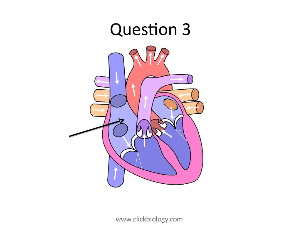
Structure of the heart without labels. cardiovascular system diagram without labels heart diagram human labels without biology anatomy bio anp physiology uploaded user. Circulatory System Respiratory System - ClipArt Best - ClipArt Best . circulatory system respiratory diagram simple heart drawing clipart cardiovascular clip clipartbest clipartmag sketch explanation cliparts heart diagram without labels heart diagram without labels Labelling the heart diagram Quiz. 14 Pictures about Labelling the heart diagram Quiz : Heart And Labels Drawing at GetDrawings | Free download, heart diagram no labels and also Free Unlabeled Heart Diagram, Download Free Clip Art, Free Clip Art on. Labelling The Heart Diagram Quiz heart labelling How the Heart Works: Diagram, Anatomy, Blood Flow - MedicineNet The heart is an amazing organ. It starts beating about 22 days after conception and continuously pumps oxygenated red blood cells and nutrient-rich blood and other compounds like platelets throughout your body to sustain the life of your organs.; Its pumping power also pushes blood through organs like the lungs to remove waste products like CO2.; This fist-sized powerhouse beats (expands and ... Heart Labeling Quiz: How Much You Know About Heart Labeling? Here is a Heart labeling quiz for you. The human heart is a vital organ for every human. The more healthy your heart is, the longer the chances you have of surviving, so you better take care of it. Take the following quiz to know how much you know about your heart. Questions and Answers 1. What is #1? 2. What is #2? 3. What is #3? 4. What is #4?
quizlet.com › 630625176 › chapter-19-the-heart-flashChapter 19: The Heart Flashcards | Quizlet •Allows heart to beat without friction, gives it room to expand and resists excessive expansion •Parietal pericardium-tough outer, fibrous layer of connective tissue-inner serous layer •Visceral pericardium (a.k.a. epicardium of heart wall)-serous lining of sac turns inward at base of heart to cover the heart surface Chambers of the Heart - Cleveland Clinic Your heart is located under your ribcage just left of your breastbone and between your lungs. The chambers within your heart are arranged in a particular way to allow blood to flow throughout your body. To remember that your atria are the "upper chambers," you can think of them as "above" your ventricles. Both atria and above begin with "a." Anatomy Lecture Midterm Flashcards | Quizlet Match each phrase to the cardiovascular system function it describes. - Vasoconstriction and vasodilation due to temperature changes. - Platelets work to plug holes in blood vessels due to trauma. - Distribution of absorbed nutrients throughout the body. - Movements of O2 to the tissues of the body - Movement of Urea to the kidneys A Diagram of the Heart and Its Functioning Explained in Detail The heart blood flow diagram (flowchart) given below will help you to understand the pathway of blood through the heart.Initial five points denotes impure or deoxygenated blood and the last five points denotes pure or oxygenated blood. 1.Different Parts of the Body. ↓. 2.Major Veins.
Heart: Anatomy and Function - Cleveland Clinic Heart. Your heart is the main organ of your cardiovascular system, a network of blood vessels that pumps blood throughout your body. It also works with other body systems to control your heart rate and blood pressure. Your family history, personal health history and lifestyle all affect how well your heart works. Appointments 800.659.7822. Free Anatomy Quiz - The Anatomy of the Heart - Quiz 1 6 - the heart : name the parts of the human heart. 7 - the muscles : Can you identify the muscles of the body? 8 - anatomical planes and directions : Do you know the language of anatomy? 9 - the spine : Test your knowledge of the bones of the spine. 10 - the skin : understand the functions of the integumentary system. heart | Structure, Function, Diagram, Anatomy, & Facts heart, organ that serves as a pump to circulate the blood. It may be a straight tube, as in spiders and annelid worms, or a somewhat more elaborate structure with one or more receiving chambers (atria) and a main pumping chamber (ventricle), as in mollusks. In fishes the heart is a folded tube, with three or four enlarged areas that correspond to the chambers in the mammalian heart. In animals ... Heart Diagram with Labels and Detailed Explanation - BYJUS The heart is made up of four chambers: The upper two chambers of the heart are called auricles. The lower two chambers of the heart are called ventricles. The heart wall is made up of three layers: The outer layer of the heart wall is called epicardium. The middle layer of the heart wall is called myocardium.
editorial « Graphic Design, Photorealistic CGI, Information Graphics, Technical Illustration ...
professional.heart.org › en › partnersFellow of the American Heart Association (FAHA) International applicants will be required to provide the same online application data as domestic applicants. Letter of Recommendation. If the candidate does not have access to a FAHA who knows their work, their letter of recommendation may be authored by the Chair or Academic Chair of their institution or by an international scientific leader.

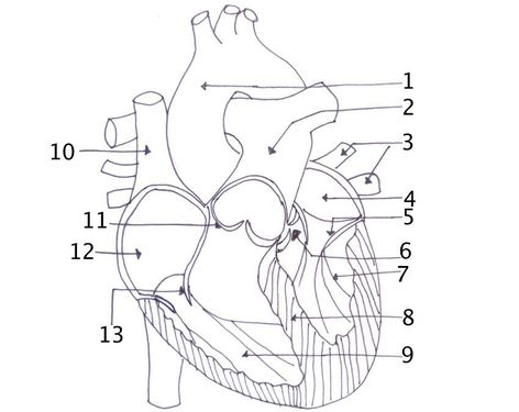
In this diagram they are showing the function of the heart as they have labels to the parts of ...
The Heart | Boundless Anatomy and Physiology | | Course Hero The heart consists of four chambers separated into two sides. Each side contains an atria which receives blood into the heart and flows it into a ventricle, which pumps the blood out of the heart. The atria and ventricle on each side of the heart are linked together by valves that prevent backflow of blood.

Free Blank Heart Diagram, Download Free Blank Heart Diagram png images, Free ClipArts on Clipart ...
Heart anatomy: Structure, valves, coronary vessels | Kenhub The heart has five surfaces: base (posterior), diaphragmatic (inferior), sternocostal (anterior), and left and right pulmonary surfaces. It also has several margins: right, left, superior, and inferior: The right margin is the small section of the right atrium that extends between the superior and inferior vena cava .
Heart structure and function - online presentation
Figuring Out Cardiac Anatomy: Your Heart - dummies In fact, your heart is situated slightly to the left of center in your chest. Figure 1: Anterior view of the heart. A thick layer of muscle tissue and a protective membrane that folds into two layers, called the pericardium or pericardial membranes, surround the heart. The heart itself is a well-organized grouping of hollow spaces.

Please label the following heart anatomy:
Human Heart (Anatomy): Diagram, Function, Chambers, Location in ... - WebMD The heart has four chambers: The right atrium receives blood from the veins and pumps it to the right ventricle. The right ventricle receives blood from the right atrium and pumps it to the lungs,...
.png)
Parts Of The Heart - ProProfs Quiz
Human Heart - Diagram and Anatomy of the Heart - Innerbody The heart contains 4 chambers: the right atrium, left atrium, right ventricle, and left ventricle. The atria are smaller than the ventricles and have thinner, less muscular walls than the ventricles. The atria act as receiving chambers for blood, so they are connected to the veins that carry blood to the heart.
Heart structure and function - online presentation

Cardiovascular System – The Internet’s Best Anatomy & Physiology Tutorial and Study Resource ...

Long Bone Label The Structure The Long Bone And Labels Label Long Bone Diagram Blank Bone ...

Label every structure on the figure of the heart: image | Study.com
Heart dissection - BIOLOGY4ISC
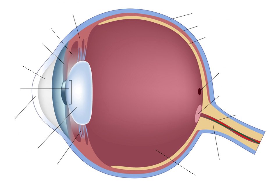
Human Eye Anatomy Quiz
Free Blank Heart Diagram, Download Free Blank Heart Diagram png images, Free ClipArts on Clipart ...

Heart label diagram
Post a Comment for "44 structure of the heart without labels"