43 diagram of the brain without labels
Brain: Function and Anatomy, Conditions, and Health Tips Brain diagram Use this interactive 3-D diagram to explore the brain. Anatomy and function Cerebrum The cerebrum is the largest part of the brain. It's divided into two halves, called hemispheres.... Somatosensory Cortex | Function, Position, Anatomy, Physiology The somatosensory cortex is a part of your brain that receives and processes sensory information from the entire body. Other names of somatosensory cortex include somesthetic area and somatic sensory area. This part of the brain is essential for receiving. sensory information from the body and processing it to initiate important movements.
en.wikipedia.org › wiki › File:Human_skeleton_frontFile:Human skeleton front en.svg - Wikipedia Restructured the image internals by adding layers, changing groupings, and adding meaningful ids and labels so that the image is easier to manipulate programmatically. Also made the labels text elements and gave them ids (it might be possible to generate : 10:17, 1 October 2007: 436 × 842 (764 KB) LadyofHats: some changes asked in FP discussion
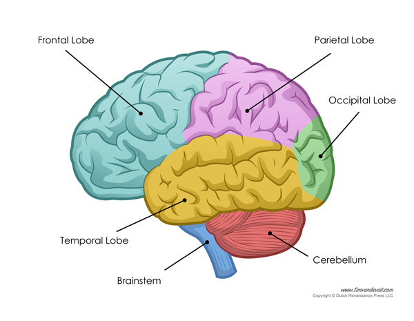
Diagram of the brain without labels
Cross-sectional anatomy of the brain - e-Anatomy - IMAIOS We created a brain atlas that is an interactive tool for studying the conventional anatomy of the normal brain based on a magnetic resonance imaging exam of the axial brain. Anatomical structures and specific areas are visible as interactive labeled images. Cross sectional anatomy: MRI of the brain Blank ear diagrams and quizzes: The fastest way to learn - Kenhub It helps you to memorize the names and their locations, which in turn will aid you to remember their functions. Below, you can download both the blank ear diagram to make some notes, and then try labeling the ear using the unlabeled ear diagram. Good luck! DOWNLOAD PDF WORKSHEET (BLANK) DOWNLOAD PDF WORKSHEET (LABELED) Ventricles of the brain: Anatomy and pathology | Kenhub It is situated in the brainstem where the ventricular surface of the rhombencephalon constitutes its floor (rhomboid fossa): inferior to the midbrain, posterior to the pons, anterior to the cerebellum and superior to the medulla oblongata. The nuclei of several cranial nerves make important impressions on the floor of the fourth ventricle.
Diagram of the brain without labels. Brain: Ultimate Guide to the Brain for AP® Psychology - Albert Resources The cerebral cortex is what you picture when you think of what a brain looks like; it is the wrinkled surface of the brain that is a layer of neurons. As we grow and learn, the neurons in our cerebral cortex grow and connects with other neurons. The cerebral cortex is made up of four lobes: Parietal, Occipital, Temporal, and Frontal. Positions and Functions of the Four Brain Lobes | MD-Health.com The brain is divided into four sections, known as lobes (as shown in the image). The frontal lobe, occipital lobe, parietal lobe, and temporal lobe have different locations and functions that support the responses and actions of the human body. Let's start by identifying where each lobe is positioned in the brain. Position of the Lobes hackaday.com › 2022/08/04 › stentrodes-a-way-toStentrodes: A Way To Insert Brain Electrodes Without Invasive ... Aug 04, 2022 · A: Diagram of a sheep brain showing the motor cortex (red), and somatotopic representations of the hindlimb (yellow), forelimb (green), head and eyes (blue), and facial muscles (purple). Anatomy of lower extremity - e-Anatomy - IMAIOS A diagram shows the various inguinal lymph nodes (lymphatic ganglia). The chapter on the innervation of the lower limb presents diagrams of the lumbosacral plexus and its main nerve branches for the lower limb (lateral cutaneous nerve of the thigh, femoral nerve, sciatic nerve and posterior cutaneous nerve of the thigh and obturator nerve).
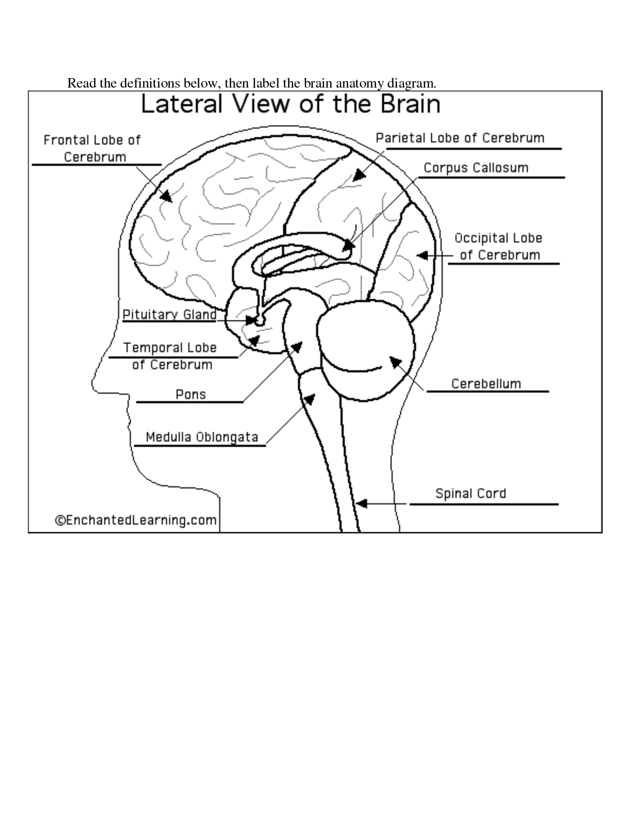
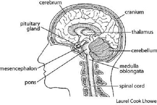
Medulla Oblongata: Anatomy, Location, and Function - Verywell Health The medulla oblongata is a tail-like structure at the base of the brain that connects the brain to the spinal cord. It carries signals from the brain to the rest of the body for essential life functions like breathing, circulation, swallowing, and digestion. While every part of the brain is important in its own way, the work of the medulla ... Free Nervous System Worksheets and Printables - Homeschool Giveaways Your kids will love seeing where the brain lobes are located under their skulls. Label the Brain Anatomy Diagram - This brain anatomy labeling worksheets is a great addition to the study of human anatomy. Human Brain Clipart and Printables - You'll find a great collection of clipart and printables showing the human brain and nerve cells. What are the 12 cranial nerves? Functions and diagram - Medical News Today The cranial nerves are a set of twelve nerves that originate in the brain. Each has a different function responsible for sense or movement. Ibai Acevedo/Stocksy. The functions of the cranial ... Whole-Brain Wiring Diagram of Oxytocin System in Adult Mice The brain was imaged as 12 × 16 × 280 tiles with 1 × 1 µm 2 x,y pixel resolution in every 50-µm z -section. We used 910-nm wavelength for two-photon excitation to excite both green (e.g., eGFP) and red signals (e.g., tdTomato). Signals were separated with 560-nm dichroic mirror and two band path filters (607/70-25 for red and 520/35- 25 for green).
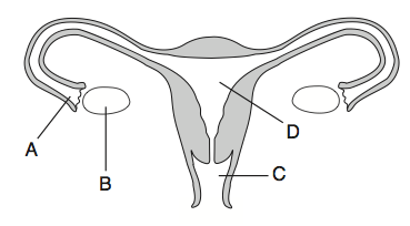
Female Anatomy: Labeled Diagrams of the Reproductive System Female anatomy refers to the internal and external structures of the reproductive and urinary systems. Reproductive anatomy aids with sexual pleasure, getting pregnant, and breastfeeding a baby. The urinary system helps rid the body of toxins through urination (peeing). The Female Reproductive System. Some people are born with internal or ... Frontal Lobe: Functions, Structure, Damage, and More - Healthline Functions of the frontal lobe. The frontal lobe controls high-level cognitive skills like: planning. self-control. memory formation. empathy. attention. It's the center for the emotions and ... How to Draw a Brain - Learn to Create a Realistic Brain Drawing The reference image that we are using within this tutorial is a diagrammatic rendition of the brain, however, it does not provide labels for each component. So try to go onto the internet and print out or have aside from a labeled brain diagram if you get confused with the names of different brain components mentioned within this tutorial. brainly.com › question › 12261308Which of these triangle pairs can be mapped to each other ... Mar 07, 2019 · A reflection creates an image that is as far behind from the reflecting line as the preimage is in front of the line. The triangle pair that can be mapped to each other using both translation and reflection across line containing AB is the first triangle pair
Human Brain Diagram - Labeled, Unlabled, and Blank
Mapping the brain to understand the mind - Knowable Magazine This closeup of a single human neuron highlights just how interconnected brain cells are. False color reveals the locations and abundance of synapses where the cell receives signals from other neurons, with excitatory inputs labeled yellow and inhibitory inputs labeled blue. CREDIT: H01 / LICHTMAN LABORATORY / GOOGLE CONNECTOMICS
9 Best Images of Brain Label Worksheet - Label the Brain Anatomy Diagram Answers, Plant Cell ...
Brain Chart Maker - 100+ stunning chart types — Vizzlo A brain chart is a fun way to visualize the things that are on your mind. A great tool to depict the functions and anatomy of the brain - ideal for school presentations. How to make a brain chart with Vizzlo? Click on the "DATA" tab to add, remove or edit records. You can also adjust the size of each area by dragging the slider.

Reflex loop👉 is a neural pathway that controls a reflex action, in higher animal most sensory ...
19 Best FREE Mind Mapping Software & Mindmap Tools in 2022 - Guru99 Add images text, labels, and annotate parts of your mind map ... Anyone can make changes in the diagram without any login using this best free mind map software. Download Link: #14) TheBrain ... Control tasks and open loops ideas in your brain. Sync your idea from your desktop, mobile device, or web browser anytime.
Brain Diagram - Cliparts.co
pressbooks.uwf.edu › medicalterminology › chapterNervous System – Medical Terminology for Healthcare Professions Figure 8.1 image description: This diagram shows a silhouette of a human highlighting the nervous system. The central nervous system is composed of the brain and spinal cord. The brain is a large mass of ridged and striated tissue within the head. The spinal cord extends down from the brain and travels through the torso, ending in the pelvis.

DotA!: Science 2nd Year 3rd Quarterly Exam Repost Brain Anatomy Diagram
› enAnatomy, medical imaging and e-learning for healthcare ... IMAIOS and selected third parties, use cookies or similar technologies, in particular for audience measurement. Cookies allow us to analyze and store information such as the characteristics of your device as well as certain personal data (e.g., IP addresses, navigation, usage or geolocation data, unique identifiers).

brain
Cerebral cortex: Structure and functions | Kenhub The cerebral cortex (cortex of the brain) is the outer grey matter layer that completely covers the surface of the two cerebral hemispheres. It is about 2 to 4 mm thick and contains an aggregation of nerve cell bodies. This layer is thrown into complex folds, with elevations called gyri and grooves known as sulci.

Brain Jack Image: กรกฎาคม 2013
14 Informative Facts, Diagram & Parts Of Human Brain For Kids - MomJunction Frontal lobe: Located at the front part of the brain, the frontal lobe is responsible for problem-solving, thinking, planning, organizing, short-term memory, movement, and motor planning, and personality characteristics. So, if you are wondering what keeps your kids organized at school or keeps your emotions under control, it is this lobe.
Brain Diagram - Cliparts.co
ppcexpo.com › blog › sankeHow to Create a Sankey Diagram in Excel Spreadsheet - PPCexpo Let’s head to the next section where you’ll learn the building blocks of the Sankey diagram. Components of a Sankey Diagram in Excel. A Sankey is a minimalist diagram that consists of the following: Nodes: This is an element linked by “Flows.” Furthermore, it represents the events in each path. Flows: Flows link the nodes. And each flow ...

What is the role of the brain in reflex action | NCERT Solutions, CBSE Sample Papers and ...
FREE Human Body Systems Labeling with Answer Sheets - Homeschool Giveaways The free respiratory system labeling sheet includes a blank diagram to fill in the trachea, bronchi, lungs, and larynx. The free nervous system labeling sheet includes blanks to label parts of the brain, spinal cord, ganglion, and nerves. The free muscular system labeling sheet includes a blank diagram to label some of the main muscles in the body.

Brain components
Brain Ventricles: Anatomy, Function, and Conditions - Verywell Health Function. Aside from cerebrospinal fluid, your brain ventricles are hollow. Their sole function is to produce and secrete cerebrospinal fluid to protect and maintain your central nervous system. CSF is constantly bathing the brain and spinal column, clearing out toxins and waste products released by nerve cells.

Bald Worm's Blog: Year 5 - Planning: The Fab Four Golden Rules
Anatomical diagrams of the brain - e-Anatomy - IMAIOS A topographical anatomy of the brain showing the different levels (encephalon, diencephalon, mesencephalon, metencephalon, pons and cerebellum, rhombencephalon and prosencephalon) as well as a diagram of the various cerebral lobes (frontal lobe, occipital, parietal, temporal, limbic and insular).

Ms Chasen's rockin' 3 & 4 psych class 2008: As requested
Brain: Atlas of human anatomy with MRI - e-Anatomy - IMAIOS Area 8 - Frontal eye fields Area 9 - Dorsolateral prefrontal cortex Areas 1-2-3 - Primary Somatosensory Cortex Arteries of brain Ascending ramus Atrium Basal vein Basilar artery Basilar part of pons Basilar plexus Basilar sulcus Body of caudate nucleus Body of fornix Brachium of inferior colliculus Brachium of superior colliculus Brain Brainstem
Ms Chasen's rockin' 3 & 4 psych class 2008: As requested
New Brain Map Charts Every Component in the Biological Universe In 2021, the project released the largest-ever map of neuronal activity and synapses in the mammalian brain. The map covered 75,000 neurons and over 500 million synaptic connections. It's a jaw-dropping scale, and the treasure trove of data is still being mined today, as scientists sleuth how form—that is, the location of neurons and ...

_Blazer_Blast: The "Why" Part of the Brain
custom-writing.org › blog › left-vs-right-brainLeft Brain vs. Right Brain: Characteristics Chart [INFOGRAPHIC] Your right hemisphere tries to call out the color, while your left side of the brain focuses on the words' meanings. If you practice a little, this funny task will become much more manageable. Practice makes perfect for every skill! That's why we have collected some useful exercises that will help you develop both your left and right brain.

Autonomic nervous system labels : Biological Science Picture Directory – Pulpbits.net
Illustrations and diagrams of the 12 pairs of cranial nerves - IMAIOS Diagrams of cranial nerves These original anatomical drawings were produced digitally, working from medical imaging sources and 3D reconstructions using Adobe Illustrator. They were then included in anatomical modules and labelled using Adobe Animate.

68 best neuro images on Pinterest | Neuroscience, The brain and Nervous system
Parts of the brain: Learn with diagrams and quizzes | Kenhub Labeled brain diagram First up, have a look at the labeled brain structures on the image below. Try to memorize the name and location of each structure, then proceed to test yourself with the blank brain diagram provided below. Labeled diagram showing the main parts of the brain Blank brain diagram (free download!)

Draw a neatly labeled diagram of Brain and write few points how it is protected. (AS5)
Ventricles of the brain: Anatomy and pathology | Kenhub It is situated in the brainstem where the ventricular surface of the rhombencephalon constitutes its floor (rhomboid fossa): inferior to the midbrain, posterior to the pons, anterior to the cerebellum and superior to the medulla oblongata. The nuclei of several cranial nerves make important impressions on the floor of the fourth ventricle.
Female Reproductive System Trivia Quiz! - ProProfs Quiz
Blank ear diagrams and quizzes: The fastest way to learn - Kenhub It helps you to memorize the names and their locations, which in turn will aid you to remember their functions. Below, you can download both the blank ear diagram to make some notes, and then try labeling the ear using the unlabeled ear diagram. Good luck! DOWNLOAD PDF WORKSHEET (BLANK) DOWNLOAD PDF WORKSHEET (LABELED)
Post a Comment for "43 diagram of the brain without labels"